Home
Research
People
Publications
Classes
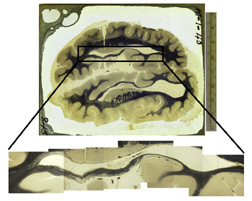
Anatomical structure and location of Von Economo neurons
 Agenesis of the corpus callosum
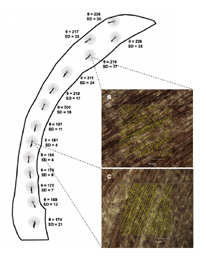
In collaboration with Ralph Adolphs and Lynn Paul, we have been investigating the anatomic structure of the brains of subjects with agenesis of the corpus callosum. This congenital failure of formation of the major pathway connecting the two halves of the brain is fairly common and is associated with abnormal social behavior and decision-making. Our data show there is a substantial reduction of all fiber systems in agenesis, and that the structure of anterior cingulate cortex is grossly abnormal in these subjects.
Neural mechanisms of humor To investigate the neural mechanisms of humor we presented cartoons to subjects in the MRI scanner and asked them to rate how funny each was. We found that activity in fronto-insular cortex (FI) is strongly related to how funny the subject rated a cartoon. We also found that verbal humor engages speech cortex, which is also activated by syntactic errors, while sight gags activate higher order visual cortical areas. Fronto-insular cortex lesions Undergraduate Corinna Zygourakis, in collaboration with Ralph Adolphs, has been investigating the deficits in social cognition that result from lesions in fronto-insular cortex. Subjects were drawn from the Iowa Patient Registry, which is a population of carefully documented neurological cases. Corinna developed a test based on film clips depicting various emotional states by first showing these clips to normal subjects to establish a baseline, then showing them to patients with FI lesions. Lesions patients had deficits in the interpretation of emotional states, particularly for the complex social emotions.
Comparative Anatomy
Aye-aye
 We performed a multi-modal analysis of tissue volume and microstructure
in the brain of the aye-aye (Daubentonia madagascariensis).
We scanned the left hemisphere of an aye-aye brain using t2-weighted
structural magnetic resonance imaging and diffusion-tensor imaging prior to
histological processing and staining for myelinated fibers. Measurements
of brain structure volumes in our specimen are consistent with those
reported in the literature: the aye-aye has a very large brain for its
body size, its visual structures (V1 and LGN) are reduced in volume,
and its olfactory lobe is increased in volume. This trade-off between
visual and olfactory reliance is a reflection of the nocturnal extractive
foraging behavior practiced by Daubentonia. Additionally, frontal
cortex volume is large in the aye-aye, a feature that could also be
related to its complex foraging behavior and increased sensorimotor
intelligence. Gross brain components appear to scale propportionally
in the aye-aye. Finally, our analysis of white matter fiber structure
in the anterior cingulum bundle demonstrates a strong correlation
between fiber spread as measured from histological sections and fiber
spread as measured from diffusion-tensor imaging.
African elephant
We performed a multi-modal analysis of tissue volume and microstructure
in the brain of the aye-aye (Daubentonia madagascariensis).
We scanned the left hemisphere of an aye-aye brain using t2-weighted
structural magnetic resonance imaging and diffusion-tensor imaging prior to
histological processing and staining for myelinated fibers. Measurements
of brain structure volumes in our specimen are consistent with those
reported in the literature: the aye-aye has a very large brain for its
body size, its visual structures (V1 and LGN) are reduced in volume,
and its olfactory lobe is increased in volume. This trade-off between
visual and olfactory reliance is a reflection of the nocturnal extractive
foraging behavior practiced by Daubentonia. Additionally, frontal
cortex volume is large in the aye-aye, a feature that could also be
related to its complex foraging behavior and increased sensorimotor
intelligence. Gross brain components appear to scale propportionally
in the aye-aye. Finally, our analysis of white matter fiber structure
in the anterior cingulum bundle demonstrates a strong correlation
between fiber spread as measured from histological sections and fiber
spread as measured from diffusion-tensor imaging.
African elephant
 We acquired magnetic resonance images (MRI) of the brain of an adult
African elephant, Loxodonta africana, in the axial and parasagittal
planes and produced anatomically labeled images. The elephant has an
unusually large and convoluted hippocampus compared to primates and
especially to cetaceans. We quantified the volume of the whole brain
and of the neocortical and cerebellar gray and white matter. The white
matter to gray matter ratio in the elephant neocortex and cerebellum
are in keeping with that expected for a brain of this size. The ratio
of neocortical gray matter volume to corpus callosum cross-sectional area
is similar in the elephant and human brains, emphasizing the difference
between terrestrial mammals and cetaceans, which have a very small corpus
callosum relative to the volume of neocortical gray matter.
We acquired magnetic resonance images (MRI) of the brain of an adult
African elephant, Loxodonta africana, in the axial and parasagittal
planes and produced anatomically labeled images. The elephant has an
unusually large and convoluted hippocampus compared to primates and
especially to cetaceans. We quantified the volume of the whole brain
and of the neocortical and cerebellar gray and white matter. The white
matter to gray matter ratio in the elephant neocortex and cerebellum
are in keeping with that expected for a brain of this size. The ratio
of neocortical gray matter volume to corpus callosum cross-sectional area
is similar in the elephant and human brains, emphasizing the difference
between terrestrial mammals and cetaceans, which have a very small corpus
callosum relative to the volume of neocortical gray matter.